General Inspection of the Abdominal Cavity and its Contents

This image shows the peritoneum following the removal of the abdominal wall.

When the abdominal wall aw is removed, what you have remaining is the transversalis fascia covering the peritoneum. It is thicker and fatty in some areas and it contains some of the blood vessels supplying the anterior abdominal wall (i.e., the inferior epigastric artery iea in this case).

After the peritoneum is opened, identify the major organs as they lie within the abdominal cavity.
In this dissection, the lower part of the peritoneum has been reflected downward in order to display the peritoneal folds in this region:
  • lateral peritoneal fold lpf produced by the inferior epigastric artery.
  • medial peritoneal fold mpf produced by the the embryonic umbilical arteries
  • median peritoneal fold mepf produced by the closed embryonic urachus which arose from the apex of the urinary bladder in the embryo.

You will frequently read that the peritoneal folds are called ligaments. In this case, only the folds formed by left-over embryonic structures are named ligaments. The embryonic urachus is the median umbilical ligament and the embryonic umbilical arteries are the lateral umbilical ligament.
Structures that should be identified at this stage are:
  • liver in the upper right quadrant of the cavity. It is separated into right rlli and left llli lobes by the falciform ligament fl.
  • the tip of the gall bladder gb hanging down under the margin of the liver
  • stomach st in the upper left quadrant
  • a small edge of the spleen sp in the upper left quadrant
  • greater omentum go covering most of the abdominal structures
  • small intestines (ileum) il in the lower right quadrant
  • sometimes the transverse colon tc can be seen through a thin portion of the greater omentum.

The free margin of the falciform ligament fl contains the remnant of the embryonic umbilical vein, now called the ligamentum teres (round ligament) lt of the liver. The paraumbilical veins run along either side of the ligament and empty into the portal vein, a clinically important connection discussed later.

At this point, you might want to take a tour of the abdominal cavity by following the peritoneum around and identifying structures as you go. Take a look at this page and follow the instructions. It would be nice if this could be done at the cadaver, but not everyone has that opportunity so you have to imagine what is going on.
When the greater omentum is pulled up and to the right side, you can see more of the small intestine si. Most of this small intestine is jejunum. The transverse colon tc is attached to the under side of the greater omentum and can also be seen.
By pulling the small intestine from the left side of the abdomen up and to the right, you can see the structures under it. The descending colon dc, a piece of the sigmoid colon sc and the upper part of the rectum rec. You can also see the root of the mesentery of the small intestine dotted line.

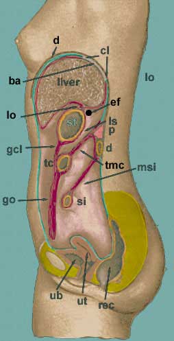
Summary of Ligaments attached to the Umbilicus


  The falciform ligament is a double fold of peritoneum which extends from the umbilicus to the antero-superior surface of the liver. In its free edge, you will see that it contains a cord-like structure which passes to the inferior border of the liver. This is the round ligament of the liver, which is formed by the remains of the left umbilical vein of the fetus. Running adjacent to the ligament are small veins the connect the paraumbilical veins around the umbilicus to the portal vein.
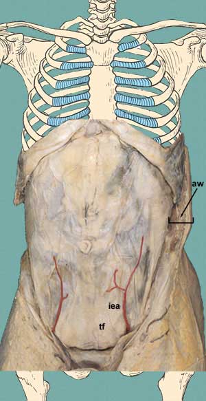
  On the deep surface of the lower abdominal wall, note that there are three cord-like structures seen through the peritoneum and extending upwards towards the umbilicus. These are the median umbilical ligament (or median umbilical fold), and the lateral umbilical ligaments (or medial umbilical folds). The median umbilical ligament extends from the tip of the bladder to the umbilicus and is the remains of the fetal urachus. The lateral umbilical ligaments arise from the pelvis as a continuation of the internal iliac artery and extend to the umbilicus. These are the obliterated parts of the fetal umbilical arteries that carried blood from the fetus back to the placenta of the mother.

Reflections of the Peritoneum

On the adjacent sagittal section of the body, you can identify the following parts of the peritoneum and its reflections. The parietal peritoneum is colored greenish blue and the visceral peritoneum is dark purple. Just imagine that you have a cadaver available. Insert your fingers between the diaphragm d and the top of the liver and push until you are stopped by a reflection of the peritoneum from the diaphragm onto the diaphragmatic surface of the liver. This reflection forms the superior limb of the coronary ligament cl. If you could push your fingers up behind the liver, you would encounter another reflection, this would be the inferior part of the coronary ligament. The two superior parts come forward and unite to become continuous with the falciform ligament.
If you continue to move your fingers along the diaphragmatic surface of the liver to its lower border and then onto the visceral surface of the organ, your fingers will be directed towards the lesser curvature of the stomach. This layer is the lesser omentum lo and extends from the visceral surface of the liver (specifically the porta hepatis region of the liver) to the lesser curvature of the stomach and then over the anterior surface of the stomach. The lesser omentum has a free margin near the gall bladder and behind this margin is the epiploic foramen ef which connects the greater peritoneal sac to the lesser peritoneal sac ls which is also known as the omental bursa.
If you place your fingers within the epiploic foramen and grasp the free margin of the lesser omentum, you will feel several structures: bile duct, hepatic artery and portal vein. We will talk about these later. If you place your fingers superiorly, you will feel the caudate lobe of the liver. If you feel posterior, you will feel the inferior vena cava and if you feel inferiorly, you will touch the first part of the duodenum.
You should constantly look at enough cross sections so that you feel comfortable with them. Remember that your left is the cross section's right since you are looking upwards towards the body. In this image, you can identify some of the peritoneal ligaments that you couldn't see on the sagittal section above. Identify the:
  • falciform ligament FL
  • lienorenal ligament LR
  • gastrolienal ligament GL
  • lesser omentum LO

Also, identify the epiploic foramen EF and the relationships of the lesser sac LS (or omental bursa).

Here is a summary of the all of the various names used to describe different parts of the peritoneal reflections:

  1. coronary ligament
  2. falciform ligament - from anterior abdominal wall to liver
  3. lesser omentum - extends from liver to lesser curvature of stomach and first part of duodenum
  4. greater omentum
  5. transverse mesocolon - from transverse colon to posterior abdominal wall
  6. mesentery of the small intestinee - from jejunum and ileum to the root of the mesentery on the posterior wall of the abdominal cavity. The root extends obliquely across the posterior abdominal wall from the beginning of the jejunum to the end of the ileum at the cecum.
  7. lienalrenal ligament - double layer of peritoneum extending from the spleen to the anterior surface of the left kidney
  8. sigmoid mesocolon - from sigmoid colon to posterior abdominal wall over the psoas muscle
  9. mesoappendix - from base of appendix to its apex

Hosted by uCoz