Large Intestine

The large intestine extends from the ileocecal junction to the anus and is about 1.5m long. On the surface, you can identify bands of longitudinal muscle fibers called taeniae coli, each about 5mm wide. There are three bands and they start at the base of the appendix and extend from the cecum to the rectum. Along the sides of the taeniae, you will find tags of peritoneum filled with fat, called epiploic appendages (or appendices epiploicae). The sacculations, called haustra, are characteristic features of the large intestine, and distinguish it from the rest of the intestinal tract.

The large intestine consists of the following parts:
  1. cecum
  2. ascending colon
  3. transverse colon
  4. descending colon
  5. sigmoid colon
  6. rectum Not seen in diagram.
  7. anal canal Not seen in diagram.
  8. anus Not seen in diagram.
There are two flexures associated with the colon:
  1. right colic flexure or hepatic flexure
  2. left colic flexure or splenic flexure


The cecum is about 6cm long and is a blind cul-de-sac which lies in the right iliac fossa. It is the part of the colon below the opening of the ileum into the colon. The cecum lies immediately behind the abdominal wall and greater omentum. There is frequently a peritoneal recess behind the cecum called the retrocecal recess and the appendix is sometimes hiding within this recess and may extend as far superiorly as the liver.

Hanging off the cecum is the vermiform appendix which opens into the cecum about 2cm below the ileocecal opening. The average length of the appendix is about 10cm and may lie in different positions. It has its own mesentery called the mesoappendix which carries the appendicular artery.

If the cecum is opened, you can identify the opening of the ileum into the cecum. This opening is surrounded by thickened muscle which forms the iliocolic valve. In this image, you can see the first part of the ascending colon with its semilunar folds.

Arterial Supply of the Colon

The colon is supplied by branches of the superior mesenteric and inferior mesenteric arteries.
    Superior mesenteric artery
  • ileocolic artery
    • superior branch that joins the right colic
    • cecal branch
    • appendicular branch
    • ileal branch
  • right colic artery
    • descending branch to join the superior branch of the ileocolic
    • ascending branch that joins the right branch of the middle colic
  • middle colic artery
    • right branch
    • left branch that joins with the ascending branch of the left colic artery
    Inferior mesenteric artery
  • left colic
    • ascending branch that joins the middle colic
    • descending branch that joins the highest sigmoid branch
  • sigmoid arteries (2-3)
    • superior sigmoid branch join the left colic
    • inferior sigmoid branch joins the superior rectal
  • superior rectal artery - not shown in the image

Venous Drainage of the Gastrointestinal Tract

The venous drainage of the gastrointestinal tract, from the lower esophagus to the upper rectum is by way of the portal venous system. This system also drains the spleen and pancreas.

The portal vein is usually described as being formed by the splenic and superior mesenteric veins. The inferior mesenteric vein then joins the splenic vein. However, there are variations to this pattern and might exist. Two of these are that the inferior mesenteric vein may join at the junction of the splenic with the superior mesenteric or the inferior mesenteric veins may join the superior mesenteric vein before it merges with the splenic. Identify the:
  • superior rectal vein
  • inferior mesenteric vein
  • splenic vein
  • superior mesenteric vein
  • esophageal veins
  • left gastric vein
  • portal vein


The numbered stars represent the areas where the portal venous system anastomoses with the caval venous system and are clinically important in portal or caval hypertension.
  1. esophageal plexus - caval drainage into azygos veins, portal drainage into the left gastric vein
  2. rectal plexus - caval drainage into middle and inferior rectal veins and then into the pudendal and internal iliac veins back to inferior vena cava, portal drainage into the superior rectal, the inferior mesenteric and the splenic
  3. paraumbilical veins - caval drainage downward to the superficial inferior epigastric vein to the femoral vein, to the external iliac, to the inferior vena cava, upward to the thoracoepigastric vein, the lateral thoracic vein, subclavian vein, superior vena cava, portal drainage through the paraumbilical vein to the portal vein.

Clinical Consideration

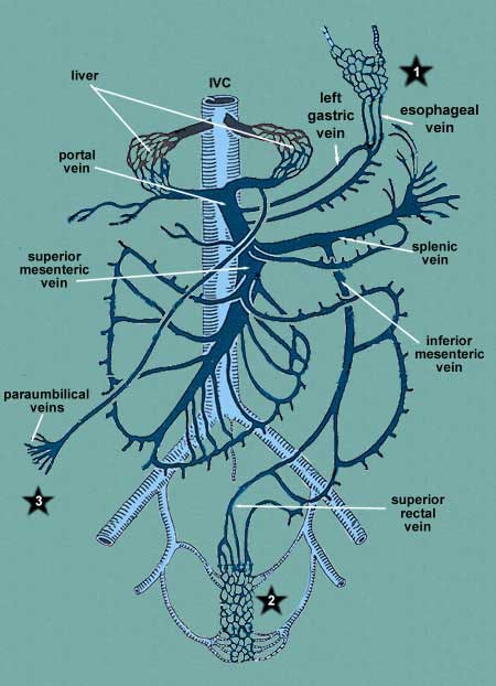
Portal obstruction. In cases of liver disease where the portal blood can no longer pass through the liver, the blood will try to get back to the heart any way it can and this usually involves the superior or inferior venae cavae. One possible cause of liver disease is chronic alcoholism. When the liver becomes impassable, it will pass backwards through the portal vein into the left gastric, paraumbilical or superior rectal. At each of these sites, the veins become enlarged and will result in other clinical signs and symptoms.

In case of the esophageal plexus (*1), esophageal varices will develop and massive hemorrhage may occur resulting in death.

In case of the rectal plexus (*2), hemorrhoids occur, resulting in pain and bleeding.

In case of the paraumbilical veins (*3), visible signs of venous enlargement and tortuosity occur on the abdomen and these are referred to the caput medusae.

Caval blockage. In cases where tumors or other pathologies compress the vena cava, the blood will utilize the above connections to return blood to the heart but this time through the caval system.
Hosted by uCoz