Liver

The liver is the largest gland of the body. It normally weighs about 1.5kg. The sharp inferior border of the liver does not normally extend below the right costal margin. If it does, it is enlarged. In order to free the liver for study, you must cut the falciform ligament, superior and inferior parts of the coronary ligament, the right and left triangular ligaments, the lesser omentum and the structures in its free margin (common bile duct, proper hepatic artery and portal vein) and the hepatic veins at the point where they empty into the inferior vena cava.
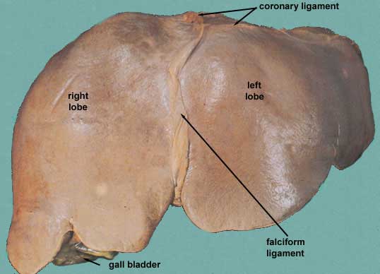
This is an anterior view of the liver. You should identify the:
  • right lobe
  • cut edge of the falciform ligament
  • left lobe
  • diverging cut edges of the superior part of the coronary ligament
  • fundus of the gall bladder
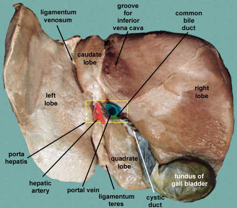
This an image of the visceral surface of the liver. Make sure you can orient yourself properly. Check out to see where the fundus of the gall bladder is located. Identify the following structures:
  • right lobe
  • fundus of the gall bladder
  • cystic duct
  • portal vein
  • hepatic arteries
  • common bile duct
  • quadrate lobe
  • ligamentum teres
  • left lobe
  • ligamentum venosum and its groove
  • caudate lobe
  • groove for the inferior vena cava and the cut hepatic veins within it
  • porta hepatis outline in yellow. The area where the arteries, ducts and portal vein enter and leave the liver.
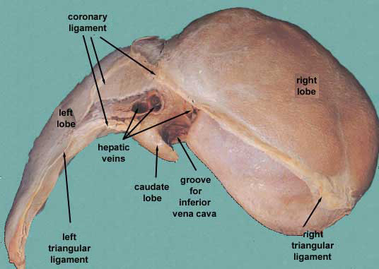
Finally we take a look at the superior aspect of the liver. This part of the liver is separated from the heart by the domes of the diaphragm. In this image, the anterior (diaphragmatic) surface of the liver is upward and the visceral surface is downward on the page. This aspect allows you to identify the:
  • right lobe
  • cut edge of the falciform ligament
  • the cut edges of the superior and inferior parts of the coronary ligament
  • the left triangular ligament
  • the right triangular ligament
  • bare area of the liver (where there is no peritoneum covering the liver
  • groove for the inferior vena cava and the hepatic veins
  • caudate lobe of the liver more or less wrapping around the groove of the inferior vena cava
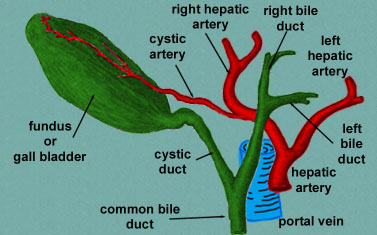
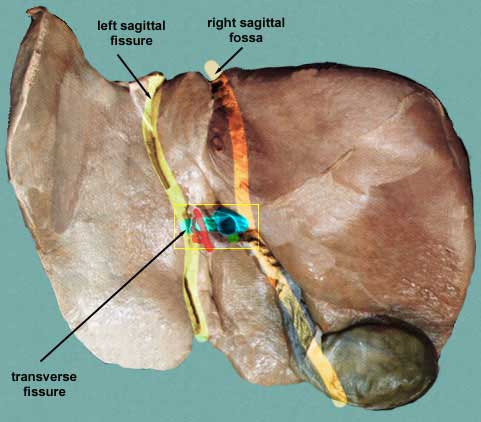
Separation of the four lobes of the liver:
  • right sagittal fossa - groove for inferior vena cava and gall bladder
  • left sagittal fissure - contains the ligamentum venosum and round ligament of liver
  • transverse fissure (also porta hepatis) - bile ducts, portal vein, hepatic arteries
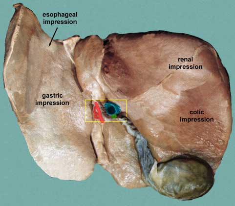
Relationship of the visceral aspect of the liver to other abdominal viscera.

Biliary System

The biliary system is made up of the ducts arising in the liver, the gall bladder and its duct and the common bile duct. Starting in the liver, the small biliary ducts converge to form the larger right and left hepatic ducts. These, in turn, join to form the common hepatic duct which joins with the cystic duct to form the common bile duct. Remember, when we studied the duodenum, that the common bile duct joins the major pancreatic duct to empty into the ampulla which then empties into the second part (descending part) of the duodenum.

The gall bladder receives bile from the liver by way of the common hepatic duct into the cystic duct. The gall bladder stores and concentrates its contents and also excretes its bile back through the cystic duct to join the common hepatic duct to become the common bile duct which then carries the bile into the duodenum.

The location of the tip of the fundus can be approximated on the surface of the abdomen at the point where the lateral edge of the rectus abdominis crosses the cartilage of the 9th rib.

Clinical Considerations

List of Items to Know

Liver
  • right lobe
  • left lobe
  • quadrate lobe
  • caudate lobe
  • falciform ligament
    • ligamentum teres of liver
  • coronary ligament
    • right triangular ligament
    • left triangular ligament
  • porta hepatis
    • common hepatic duct
    • portal vein
    • proper hepatic artery
      • left hepatic
      • right hepatic
        • cystic
    • gall bladder
      • fundus
      • cystic duct
    • ligamentum venosum or groove for the ligament
Celiac Trunk
  • common hepatic artery
    • proper hepatic artery
      • left hepatic
      • right hepatic
        • cystic artery


Hosted by uCoz