Structures of the Posterior Abdomen

Peritoneum

After the gastrointestinal tract is removed, what you have left in the posterior abdomen are the cut edges of the peritoneum and the remaining peritoneum that covers the "retroperitoneal" structures.

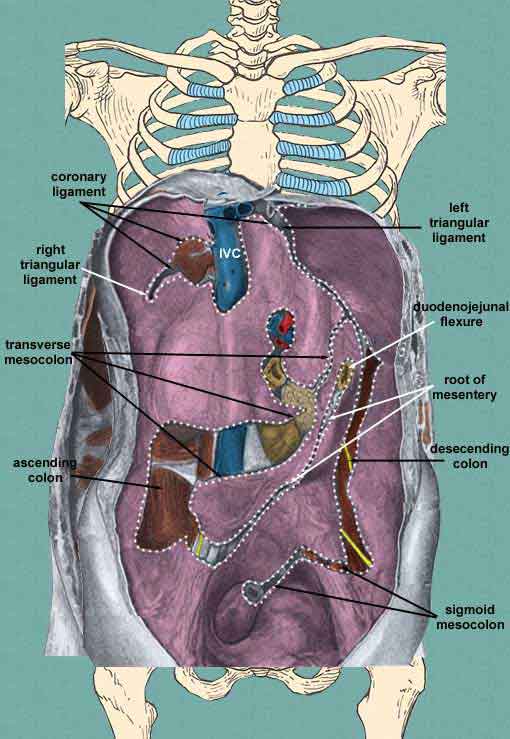
It is worth while to take a look at this type of image to appreciate just how the various mesenteries are reflected from the posterior abdominal wall as well as the diaphragm.

Identify the:
  • coronary ligament and its triangular parts
  • cut edges of the transverse mesocolon
  • area where the ascending colon used to be
  • cut edges of the sigmoid mesocolon
  • space where descending colon used to be
  • root of the mesentery
  • duodenojejunal flexure

Retroperitoneal Structures of the Abdomen

After the mesentery has been cleaned from the posterior abdominal wall, you can see the true retroperitoneal structures of the abdomen. These are the great vessels and their branches, sympathetics, kidneys and their ureters, and suprarenal glands.

You should be able to see the following structures:
  • inferior vena cava (IVC)
    • testicular (or ovarian)
  • aorta
    • celiac trunk
    • superior mesenteric artery
    • inferior mesenteric artery
    • external iliac
    • internal iliac
    • testicular (or ovarian)
  • lumbar sympathetic chain
  • celiac ganglia
  • kidney
    • ureter
  • suprarenal gland

Structure of the Kidney

When the kidney is opened, you can see the following structures:
  • cortex
  • medulla
    • renal columns
    • renal pyramid
  • interlobar arteries
  • renal papilla
  • minor calyx
  • major calyx
  • renal pelvis
  • ureter

Muscles and Nerves of the Posterior Abdominal Wall

Finally, with the kidneys and their related structures removed, you can see the muscles that make up the posterior abdominal wall and the branches of the the lower thoracic and lumbar nerves.

You should be able to point out the:
  • diaphragm
    • right and left parts of the diaphragm
    • right crus
    • left crus
    • medial arcuate ligament arches over the sympathetic trunk as it enters the abdomen and the upper fibers of the psoas muscle.
    • lateral arcuate ligament arches over the free tip of the twelfth rib and the subcostal nerve (T12)
    • median arcuate ligament arches over the aorta and the cysterna chyli (a lymphatic sac that continues into the thorax as the thoracic duct.
  • quadratus lumborum
  • psoas major
  • psoas minor (frequently absent)
  • iliacus
  • nerves
    • sympathetic trunk
    • hypogastric plexus
    • T12 - subcostal
    • L1
      • iliohypogastric
      • ilioinguinal
    • genitofemoral - lies on top of the psoas major muscle
    • lateral femoral cutaneous
    • femoral - lateral to the psoas major
    • obturator - medial to the psoas major

Openings of the Diaphragm

The diaphragm has several structures passing through it and these openings are found at different vertebral levels as follows:

Lumbosacral Plexus

The nerves of the posterior abdominal wall are branches of the lumbosacral plexus. This plexus is shown in the adjacent image.

You should be able to identify the roots of the plexus:
  • L1
  • L2
  • L3
  • L4
  • L5

L1 gives rise to the iliohypogastric and ilioinguinal nerves.

L1 + L2 gives rise to the genitofemoral nerve

L2 + L3 gives rise to the lateral femoral cutaneous

L2 + L3 + L4 give rise to the femoral and obturator nerves

L4 + L5 give rise to the lumbosacral trunk which joins sacral nerves to form the sacral plexus.

Arteries and Veins of the Posterior Abdomen

Immediately after the aorta enters the abdomen under the median arcuate ligament, it gives rise to its first paired branches, the inferior phrenic arteries.

You can break up the branches of the aorta into paired and unpaired branches. The unpaired branches are:
  • celiac trunk
    • splenic - pancreas, spleen, fundus of stomach, left part of greater curvature of stomach
    • left gastric - esophagus, lesser curvature of the stomach
    • common hepatic - liver, gall bladder, right side of lesser curvature of stomach, pancreas, duodenum, right side of greater curvature of stomach
  • superior mesenteric - pancreas, duodenum, jejunum, ileum, ascending colon, transverse colon
  • inferior mesenteric - descending colon, sigmoid colon, upper rectum
  • median sacral


The paired branches supply the organs and muscular walls of the abdomen as well as pelvic structures and the lower limb. They are:
  • inferior phrenic
    • superior phrenic
  • middle suprarenal
  • renal
    • inferior phrenic
  • lumbar
  • common iliacs
    • external iliac
      • deep circumflex iliac
      • inferior epigastric
    • internal iliac - studied with pelvis
The veins all drain into the inferior vena cava and thus back to the heart. The major veins are:
    • external iliac
    • internal iliac
  • common iliac
  • lumbar veins
  • left renal vein
    • left testicular or ovarian vein
    • suprarenal vein
  • right renal vein
    • suprarenal vein
  • hepatic veins
  • inferior vena cava


Hosted by uCoz